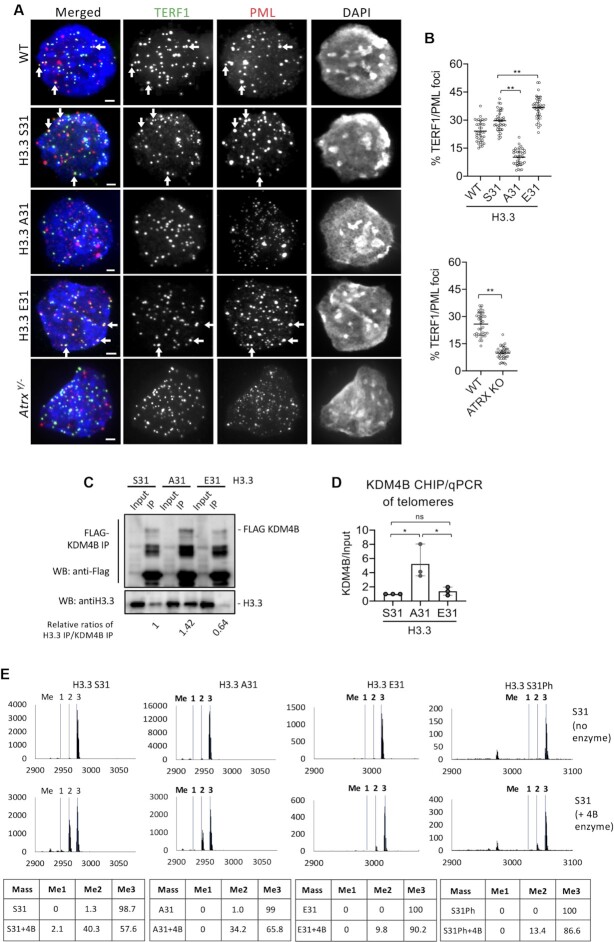
Figure 2.
H3.3 S31Ph regulates telomeric localisation to PML bodies and KDM4B activity in mouse ES cells. (A) Immunostaining of PML (red) and TERF1 (green; marker for telomere) showing co-localisation of PML and TERF1 (shown by arrows) in wildtype (WT), H3.3 S31, E31 and Atrx–/ymouse ES cell lines, but the co-localised signal was greatly reduced in H3.3 A31 and Atrx–/y mouse ES cell lines. Scale bars: 4 μm. (B) Percentages (mean ± SEM, n = 3, 16 nuclei were analysed in each experiment) of co-localized PML/TERF1 foci in wildtype (WT), H3.3 S31, A31, E31 and Atrx–/y mouse ES cell lines are shown. (C) Protein immunoprecipitation with an anti-Flag antibody in H3.3 S31, A31 and E31 mouse ES cell lines expressing Flag-tagged KDM4B, followed by western blot analysis with anti-Flag and H3.3 antibodies, respectively. The changes in H3.3/KDM4B binding affinities were presented as the relative ratios of H3.3 and KDM4B immunoprecipitated (H3.3 Ip/KDM4B Ip) in WT, A31 and E31 cell lines, respectively. (D) ChIP-qPCR analyses (mean ± SEM, n = 3) of KDM4B in H3.3 S31, A31 and E31 mouse ES cell lines, showing increased KDM4B binding (KDM4B/Input) at the telomere. (E) Mass spectrometry analysis of in vitro KDM4B histone demethylase assays. KDM4B recombinant protein was incubated with either H3.3 S31, A31, E31 or S31Ph K36me3 peptides. Dashed lines indicate expected masses for K36 me0, me1, me2 and me3. (B, D) P-values calculated using Student's t-test (** P< 0.05; * P< 0.1; ns, non-significant).

