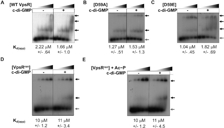
Figure 3.
DNA binding to PvpsL measured by EMSAs. Representative gels showing the retardation of the 32P-labeled DNA harboring −97 to +113 of PvpsL with increasing amounts of the indicated VpsR (concentrations of 0, 0.2, 0.4, 0.8 and 2 μM) either in the absence or in the presence of 50 μM c-di-GMP, as indicated. Black arrows indicate retarded complexes, while gray arrow indicates free DNA. Apparent DNA-binding dissociation constants (Kd(app)), determined from at least three replicate EMSAs, were calculated as the concentration of VpsR needed for the loss of 50% of the free DNA. WT VpsR and the variants (A–C) were present in Pi/high-salt buffer, while VpsRren (D and E) was present in the Tris/low-salt buffer. Consequently, the determined Kd(app) values determined in panels (A–C) are not comparable to those obtained in panels (D) and (E).

