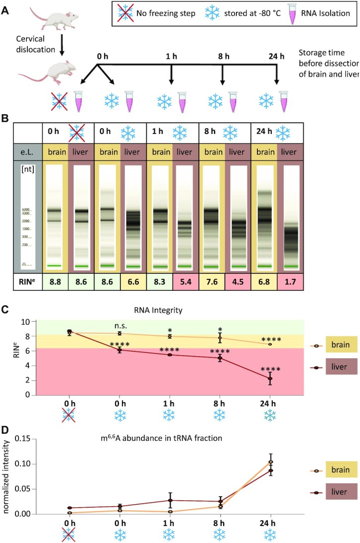
Figure 3.

In cellulo degradation of total RNA after organismal death. (A) Workflow: mice were sacrificed by cervical dislocation. RNA from liver and brain tissue was isolated at different time points post mortem. Additionally, the effect of shock freezing was assessed from RNA isolated immediately after dissection. (B) Representative TapeStation Profiles of total RNA from brain (light brown) and liver (dark brown), respectively, at the time points indicated in A. (C) RNA integrity (RINe) values of liver and brain RNA plotted over time (mean ± SD, n = 4 biological replicates, n.s. = not significant, *P< 0.05, ****P< 0.0001, unpaired t-test, compared to 0 h without freezing). (D) Relative Quantification of the rRNA marker m6,6A by LC–MS/MS shows elevated m6,6A levels in tRNA fraction of liver and brain RNA in a time-depentend manner. Mean ± SD, n = 4 biological replicates.
