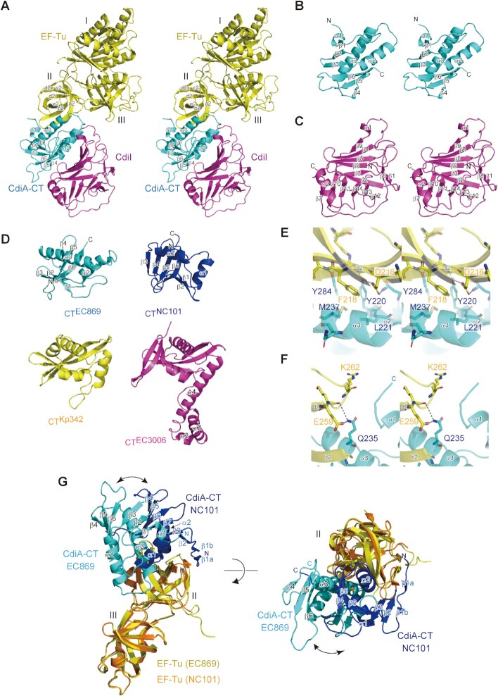
Figure 6.
Structure of CdiA–CT:CdiIEC869 complexed with Tu. (A) Stereoview of the structure of the CdiA–CT:CdiI:Tu complex. CdiA–CTEC869, CdiIEC869 and Tu are colored cyan, magenta, and yellow, respectively. The modeled CdiA–CTEC869 contains residues 175–285 (numbered from Val1 of the VENN peptide motif, Supplementary Figure S13A), and the modeled CdiIEC869 contains residues 3–179. The modeled Tu contains residues 10–393. (B) Stereoview of CdiA–CTEC869. (C) Stereoview of CdiIEC869. (D) CdiA–CTEC869 adopts the BECR fold as observed in other CdiA–CTs targeting the 3′-acceptor region of tRNAs. CdiA–CTNC101 (blue) from E. coli NC101 (14), CdiA–CTKp342 (yellow) from Klebsiella pneumoniae, and CdiA–CTEC3006 (magenta) from E. coli 3006 (16). (E), (F) Interaction between CdiA–CT (cyan) and domain II of Tu (yellow). (G) Superimposition of the structure of CdiA–CTEC869:Tu onto that of CdiA–CTNC101:Tu (14). For clarity, the structures of domain I of Tu in both complex structures are omitted. CdiA–CTEC869 and domains II/III of Tu in the structure of CdiA–CTEC869:Tu are colored cyan and yellow, respectively. CdiA–CTNC101 and domains II/III of Tu in the structure of CdiA–CTNC101:Tu are colored blue and orange, respectively.

