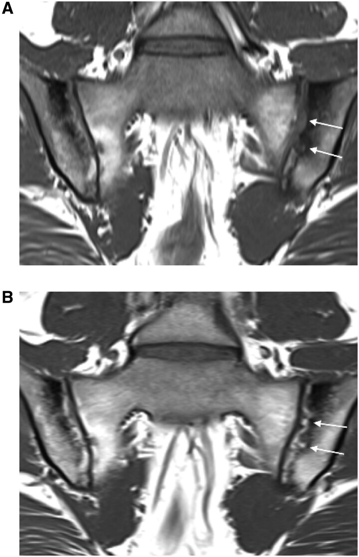
Fig. 2.
Illustrative T1W MRI scans at (A) baseline and (B) week 12 from a patient who received filgotinib
In (A) the arrows point to an extensive erosion of the left iliac bone on the T1W MRI scan. In (B) the arrows demonstrate the appearance of bright tissue filling in the cavity of the erosion, bordered by an irregular dark band. This is the characteristic appearance of backfill.

