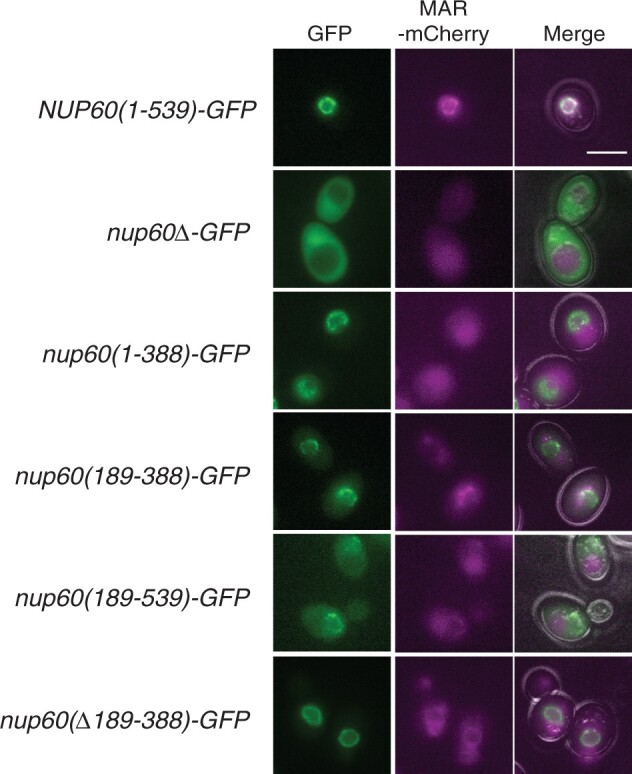
Fig. 6.

Effect of Nup60 truncations on the localization of Nup60-GFP and MAR-mCherry to the nuclear envelope. Representative images of cells expressing wild-type MAR-mCherry and GFP fusions to the Nup60 truncations illustrated in Fig. 4 are shown. The right column is a merged image of the GFP, mCherry, and bright-field channels. All strains are isogenic to the NUP60(1–539)-GFP strain: (1–539) (SBY6254), nup60Δ (SBY6338), nup60(1–388) (SBY6344), nup60(189–388) (SBY6341), nup60(189–539) (SBY6347), and nup60(Δ189–388) (SBY6350). The intensity of the NUP60(1–539) MAR-mCherry panel was adjusted to highlight MAR-mCherry’s localization to the NE. The scale bar represents 5 µm.
