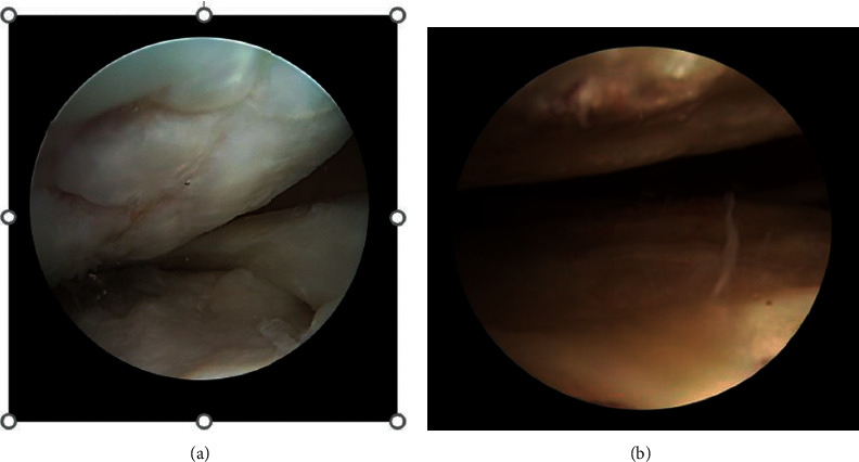
Figure 7.

Intraoperative second-look arthroscopic images of cartilage repair sites (after 12 months). (a)The original chondral lesion is filled with fibrocartilagineous tissue after microfractures + PRP (platelet-rich plasma) injection. (b) A hyaline-like cartilage filling the chondral defect is detectable after microfractures + PRP + AD-MSC (adipose-derived mesenchymal stem cells) treatment.
