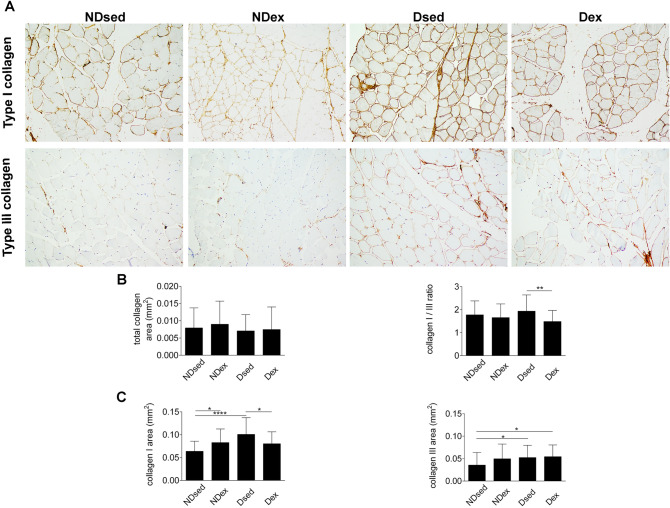
Figure 3.
Immunohistochemical sections of rat rectus abdominis muscle (RAM), obtained from CellSens Dimension (Olympus Corporation®) Version 1.16 image analysis software—(https://www.olympus-lifescience.com/en/software/cellsens/). (A) RAM samples were taken from non-diabetic sedentary (NDsed), non-diabetic exercised (NDex), diabetic sedentary (Dsed) and diabetic exercised (Dex) rats as described in Materials and methods. RAM samples were stained with Picrosirius red and immunohistochemistry using antibodies against type I and III collagen on RAM fibers. (B) Picrosirius red staining for total collagen area and immunohistochemistry for type I/III ratio. (C) Immunohistochemistry for type I and III collagen area as in (A). Values are means ± S.D. (n = 5 animals/group. *p < 0.05, **p < 0.01, ***p < 0.001 and ****p < 0.0001. Scale bar: 50 µm. Magnification: ×20.

