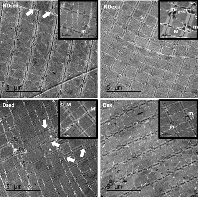
Figure 4.

Electron micrographs of rectus abdominis muscle (RAM). Samples of RAM were obtained from non-diabetic sedentary (NDsed), non-diabetic exercised (NDex), diabetic sedentary (Dsed), and diabetic exercised (Dex) rats. Magnifications (20.000 x) show a detailled area of the micrograph. The micrographs show disorganized Z lines (white arrows), sarcomeres disruption areas (asterisk), intermyofibrillar mitochondria (m), myelin figures (M), organized triads (t) and an increase in collagen deposition ( +). Scale bar: 5 µm.
