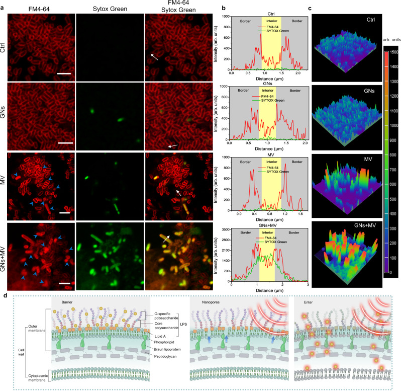
Fig. 6. Mechanism of GNs collaboration with MV to kill E. coli.
a Fluorescent localization of the interactions between GNs, MV, and E. coli. The fluorescent dyes FM4-64 to stain the OM (red), and SYTOX Green to show membrane permeabilization (green). Scale bar, 5 μm. b Fluorescence intensity along the white arrows in a. c Intensity surface plot of different groups in a. d Schematic diagram of MV and GNs synergistically killing bacteria. GNs are represented by yellow spheres. Source data are provided as a Source Data file.

