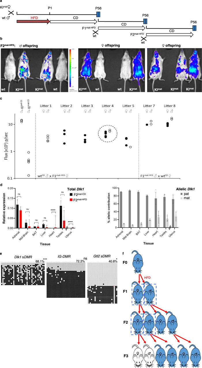
Fig. 5. Exposure-induced changes to Dlk1 imprinting are transmitted to F2 offspring.
a Schematic for generational studies following HFD exposure. Gestationally exposed animals (Dlk1-FLucLacZ F1mat-HFD) were bred with wt (CD-fed) mates, maintained on CD, and F2 and F3 offspring examined. b BL signal (blue) in F2 offspring (F2mat-HFD) derived from F1 HFD-exposed females. Signal was variable and ectopic. c Abdominal BL signal in P56 F2mat-HFD males (open-circles) and females (filled-circles), from six F1mat-HFD females and wtCD males (litters 1–5, no litter from female 6) or two F1mat-HFD males and wtCD females (litters 7–8). KImat-CD and KIpat-CD signal shown for comparison. Litter 4 is represented in (b). Source data are provided as a Source Data file. d Dlk1 expression (QRT-PCR, left) in tissues from P56 males (uterus from females) whose mothers were exposed in utero to CD (F2mat-CD, black) or HFD (F2mat-HFD, red). Expression normalised to β-Actin, 18S and Hprt (bars show geometric mean with geometric SD; N = 4 + 4 individual mice; Two-way ANOVA on delta-Ct values (Tissue p < 0.0001, Diet p = 0.002, Interaction p < 0.0001); Holm-Šídák’s multiple comparisons follow-up test for diet in each tissue: ****padj < 0.0001, ns=not significant). Allelic Dlk1 analysis in F2mat-HFD mice (right) showed reduced paternal (dark grey) versus maternal (light grey) expression bias, compared to control conditions (bars indicate mean allelic contribution ±SD; N = 4 + 4 individual mice). Source data are provided as a Source Data file. e Bisulphite analysis in male P56 F2mat-HFD liver showed Dlk1 sDMR hyper-methylation, increased IG-DMR methylation (padj = 0.078) and slightly reduced Gtl2 sDMR methylation, compared to F1mat-CD (Fig. 4a). Closed circles: methylated CpG, open circles: un-methylated CpG. Rows show individual clones from a representative individual, percentages indicate total methylation from two animals. (Kolmogorov-Smirnov test comparing clonal methylation levels, using Holm-Šídák’s correction for multiple comparisons: *padj = 0.025, ***padj = 0.0001, ns=not significant). Source data are provided as a Source Data file. f Summary of altered Dlk1 expression following gestational HFD. Dlk1 is silent (white) when transmitted maternally and expressed (blue) when transmitted paternally. Gestational HFD exposure provokes LOI in F1 offspring (blue, box). F1 females transmit altered Dlk1 expression to F2 offspring (blue, box), whereas F1 males and F2 females transmit Dlk1 appropriately.

