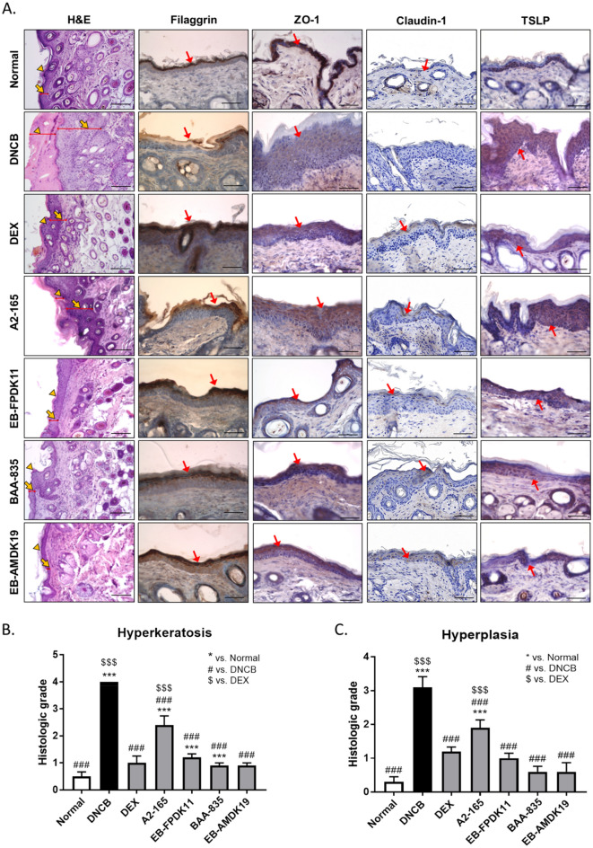
Figure 4.
EB-FPDK11 and EB-AMDK19 enhance skin barrier proteins in AD-like lesion in NC/Nga mice. (A) The first lane: H&E staining of dorsal skin lesions. The yellow arrowheads indicate hyperplasia, and the yellow arrows indicate hyperkeratosis. The 2nd–4th lane: Immunohistochemical staining of filaggrin, ZO-1, claudin-1, and TSLP, respectively in dorsal skin lesions in DNCB-induced NC/Nga mice. Dark brown regions (the red arrows) indicate positively stained cells. Scale bar, 50 µm. (B, C) The graphs represented as (B) Hyperkeratosis (C) Hyperplasia, histological grades were scored as follows: 0, normal thickness; 1, two times normal thickness; 2, three times normal thickness; 3, four times normal thickness; or 4, greater than four times normal thickness. Data are presented as mean ± SEM of changes in values. *P < 0.05; **P < 0.01; ***P < 0.001; *versus normal group; #versus DNCB group; $versus DEX group.

