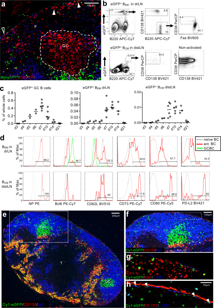
Fig. 1. Appearance of antigen-activated memory-like B cells in drLN and distLN.
a Location of B1-8i/k−/−/Blimp1GFP/Cdt1mKO2 B cells in drLN 6 d after immunization. Antigen-specific B cells in the G0/G1 phase of cell cycle (red) inside the GC (dashed line) and in the follicle (F) close to the SCS (arrow heads). Interfollicular region (open triangle). Blimp-1+ PCs are eGFP (green). Hoechst33342-labeled naïve T cells (blue). Scale bar: 100 μm. Representative image of 3 lymph nodes. b Gating of eGFP+ BEM in drLN and BCM and distLN 8 d after immunization of recipients of NP-specific Cγ1Cre QM mTmG B cells. c Kinetic of eGFP+ B cell appearance in drLN and distLN. Data merged from two independent experiments (n = 2–3). Each datapoint represents one animal. d Memory B cell typical markers on BEM and BCM in drLN and distLN. e drLN of recipient of QM Cγ1Cre mTmG cells 8 d after immunization. T zone (T). f Enlargement of box in e showing the eGFP+ BEM close to the subcapsular sinus, g Ki-67 expression in BEM, h same area showing BEM location below the LYVE1+ ER-TR7-ve SCS floor endothelium and inside the SCS (arrowheads). Image is a representative of three lymph nodes.

