. 2019 Oct 3;9(53):31186–31195. doi: 10.1039/c9ra01792e

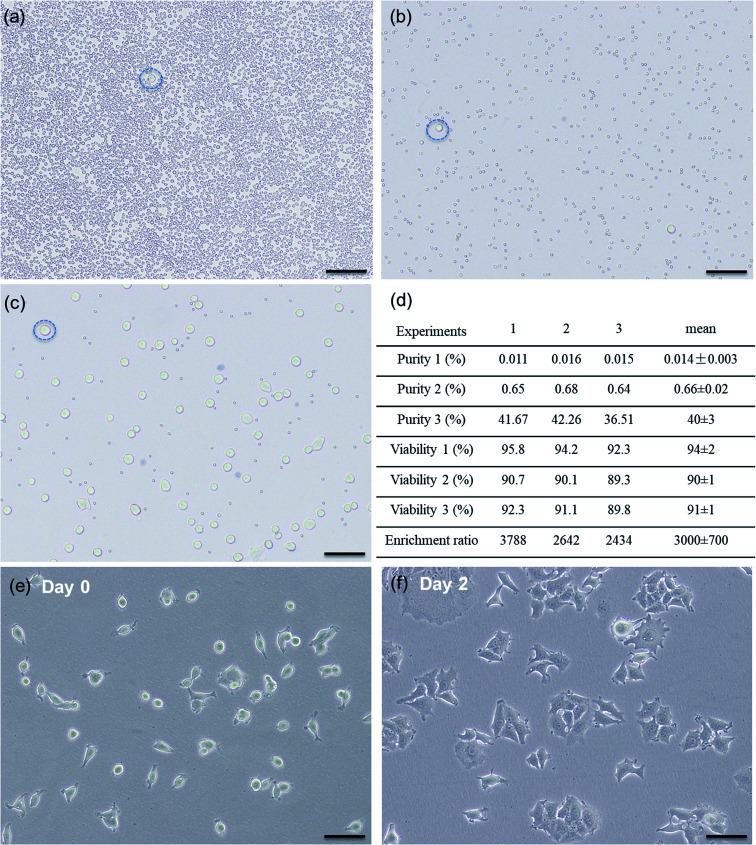
Fig. 5. Representative microscopic photographs of cell samples collected from (a) input inlet of inertial sorting device, (b) target outlet of inertial sorting device, and (c) target outlet of acoustic sorting device. A representative cancer cell in each sample is indicated by a blue dashed circle. Scale bar is 100 μm. (d) Experiments 1–3 refer to the three repeated experiments under the same sorting conditions. Purity 1 and viability 1 refer to the purity and viability of cancer cells from sample (a). Purity 2 and viability 2 refer to the purity and viability of cancer cells from sample (b). Purity 3 and viability 3 refer to the purity and viability of cancer cells from sample (c). (e) and (f) show proliferation of cancer cells collected from the final sorting output.

Fig. 5