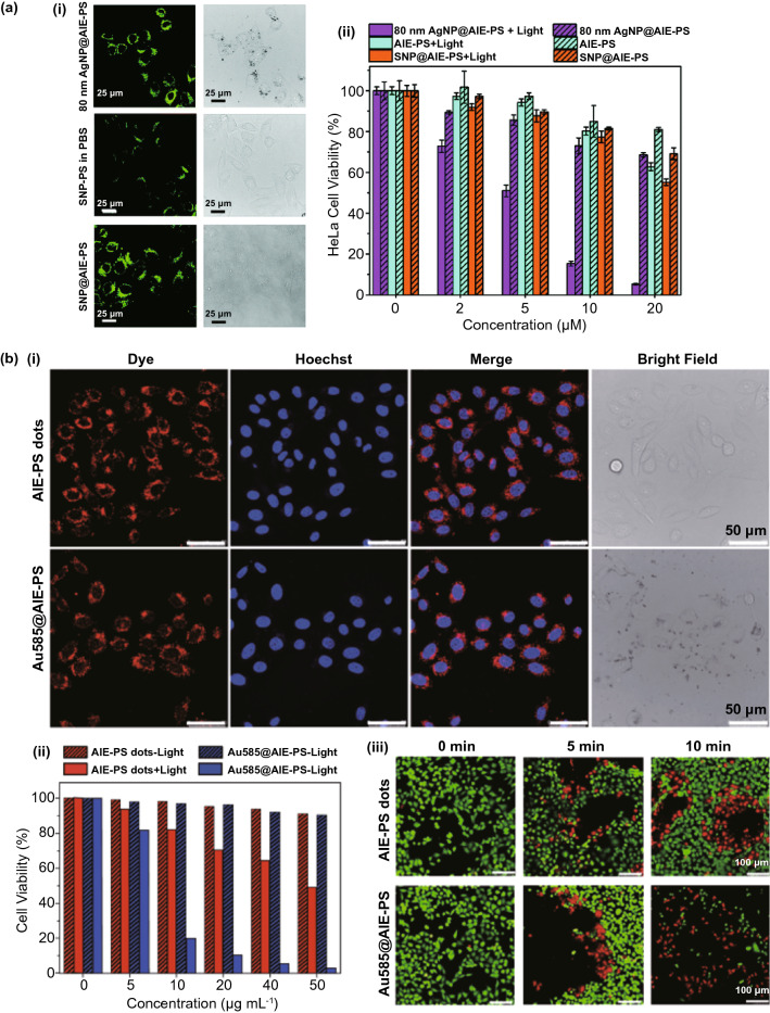
Fig. 13.
a (i) Confocal fluorescence imaging and bright field images of HeLa cells incubated with 80 nm AgNP@AIE-PS (top row) and AIE-PS dots (bottom row) in physiological buffer solution (PBS), respectively. Silica nanoparticles (SNP) @AIE-PS is used as the control samples, (ii) cytotoxicity of the same samples in a under white light illumination and at darkness against HeLa cells, reproduced from Ref. [22] with permission from Royal Society of Chemistry, b (i) Confocal fluorescence imaging and bright field images of HeLa cells incubated with AIE-Ps dots and Au585@AIE-PS dots samples. The nuclei were stained with Hoechst dye., (ii) Cell viability of HeLa cells after incubation with different concentrations of AIE-PS and Au585@AIE-PS samples with and without white light illumination. (iii) Live/dead cell staining of HeLa cells before (0 min) and after light treatment for 5 and 10 min, respectively, reproduced from Ref. [21] with permission from Springer-Nature

