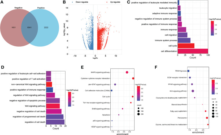
Figure 3.
Global m6A modification changes in the normal livers and ALV-J induced tumor livers. (A) Venn diagram of m6A methylation sites identified in mRNAs from two groups. (B) The Volcano plot showed the identification of significant hyper- and hypomethylated m6A peaks. (C, D) GO enrichment analysis of biological processes involved in hypermethylated (C) and hypomethylated (D) m6A genes in ALV-J infected positive group compared with negative group. (E, F) Pathway analysis of hypermethylated (E) and hypomethylated (F) m6A genes in mRNAs of ALV-J infected positive group compared to negative group. Negative represents chickens without ALV-J infection (normal livers); Positive represents chickens with ALV-J infection (tumor livers).

