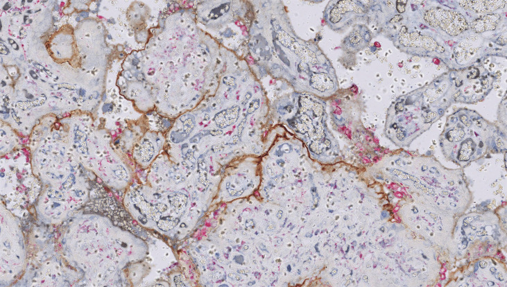
Figure 4.
Chronic histiocytic intervillositis (CHI). Dual staining demonstrates aggregates of CD68+ histiocytes in the intervillous space (pink) and linear deposition of C4d along the microvillous border (brown) in a case requiring delivery by emergency Caesarean section for severe fetal growth restriction with abnormal umbilical artery Dopplers at 24 weeks’ gestation. Histology also showed extensive perivillous fibrin deposition.

