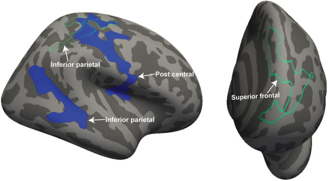
FIGURE 2.
For ME/CFS and HC, significant clusters from interaction-with-group regressions for 2 clinical regressors (“Fatigue” and “HRV”). The volume and thickness cluster of the post central gyrus and inferior parietal was observed in the left hemisphere when regressed with “Fatigue” (left side). The thickness cluster of the superior frontal gyrus was detected at the left hemisphere when regressed with “HRV” (right side). The volume is represented with filled blue color whereas thickness is represented by unfilled green color. Significant volume and thickness clusters were overlaid on the inflated brain (left and right hemisphere) available in the FreeSurfer.

