Figure 2.
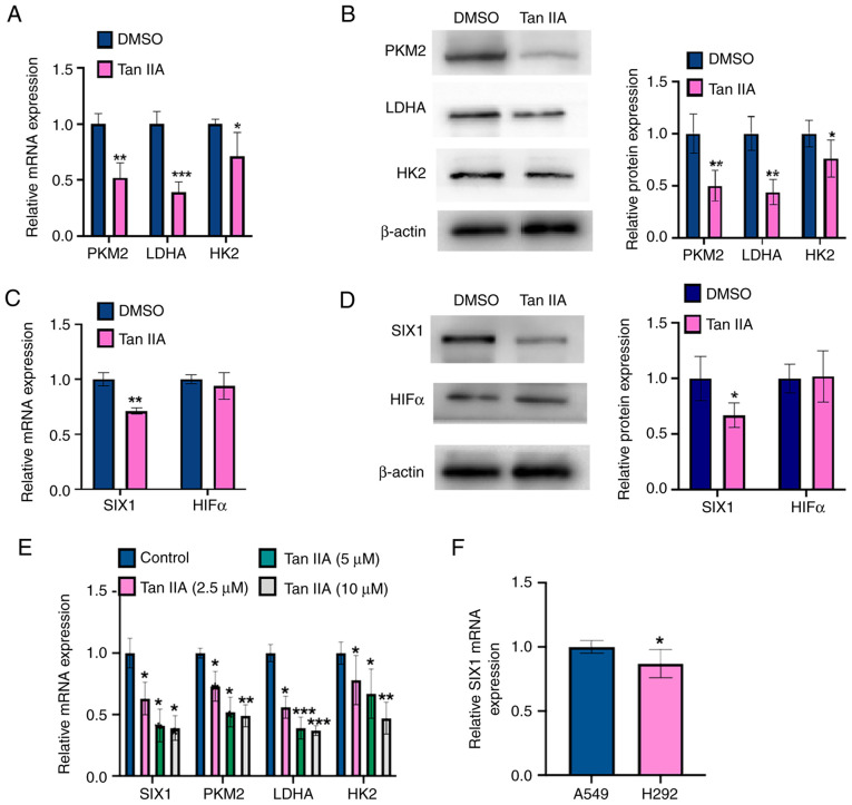
Tan IIA downregulates SIX1 expression in NSCLC cells. (A) A549 cells were treated with 5 µM Tan IIA for 48 h. The mRNA levels of PKM2, LDHA and HK2 were detected by RT-qPCR. *P<0.05, **P<0.01 and ***P<0.001 vs. DMSO. (B) A549 cells were treated as in (A), and the protein levels of PKM2, LDHA and HK2 were detected by western blot analysis. The right panel shows the densitometric analysis of three independent experiments. *P<0.05 and **P<0.01 vs. DMSO. (C) A549 cells were treated as in (A), and the mRNA levels of SIX1 and HIF1α were detected by RT-qPCR. **P<0.01 vs. DMSO. (D) A549 cells were treated as in (A), and the protein levels of SIX1 and HIF1α were detected by western blot analysis. The right panel shows the densitometric analysis of three independent experiments. *P<0.05 vs. DMSO. (E) A549 cells were treated with Tan IIA (2.5, 5 and 10 µM) and mRNA levels of PKM2, LDHA, HK2 and SIX1 were detected by RT-qPCR. *P<0.05, **P<0.01 and ***P<0.001 vs. DMSO. (F) RT-qPCR was used to detect the mRNA level of SIX1 in A549 and H292 cell lines. *P<0.05 vs. A549 cell line. Tan IIA, Tanshinone IIA; SIX1, sine oculis homeobox homolog 1; PKM2, pyruvate kinase subtype M2; HK2, hexokinases 2; LDHA, lactate dehydrogenase A; HIF1α, hypoxia inducible factor 1α; RT-qPCR, reverse transcription-quantitative PCR.

