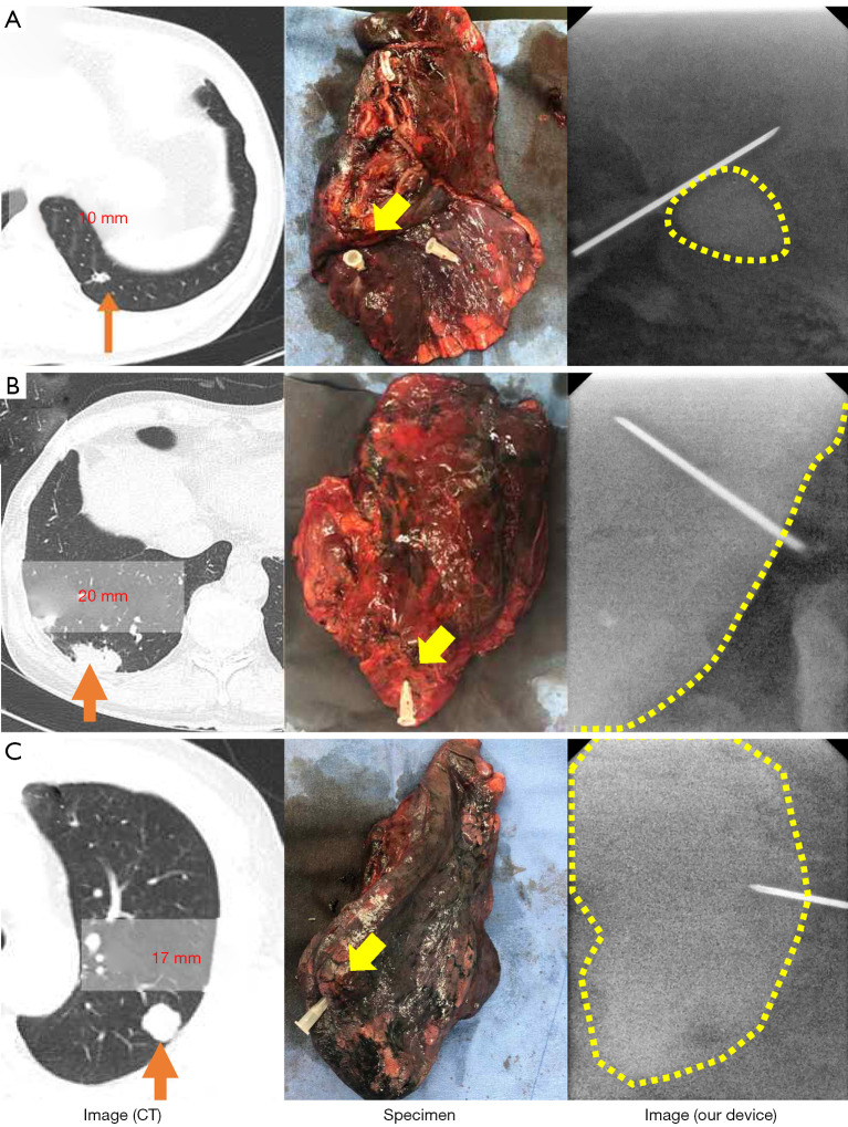
Figure 4.
X-ray image of human lung cancer specimens acquired using computed tomography (CT) and our device without injecting radiocontrast agent. (A) Case of a 55-year-old male patient with a 10-mm-sized solid lesion in the left lower lobe of the lung with suspected lung metastasis from colorectal cancer. (B) Case of a 68-year-old male patient with 20-mm-sized primary early lung cancer in the right lower lobe. (C) Case of a 68-year-old male patient with 17-mm-sized primary lung cancer in the left upper lobe. Although no radiopaque contrast agents such as lipiodol were injected before or during the surgery, the X-ray images obtained using the proposed device clearly show the needle-marked solid lesion.

