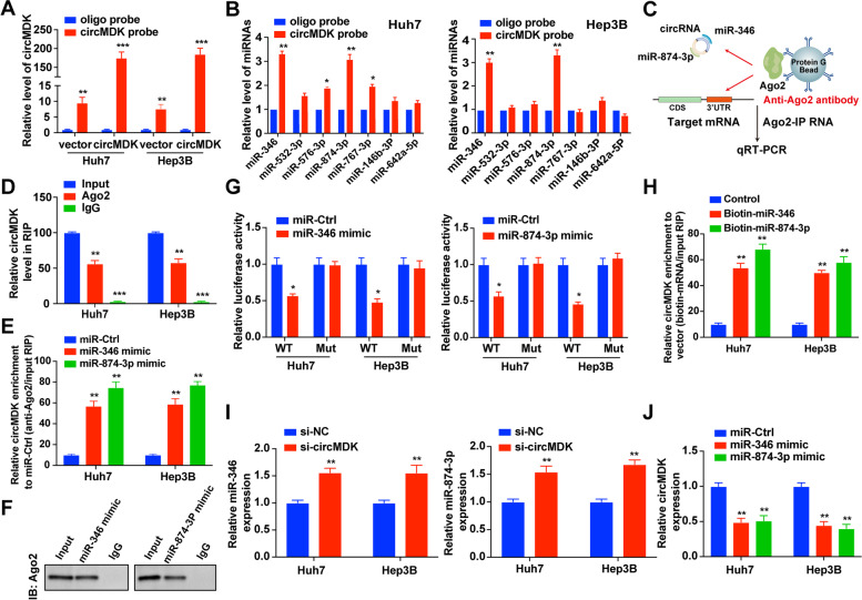
Fig. 4.
CircMDK acts as a ceRNA and sponges miR-346 and miR-874-3p. A Lysates from Huh7 and Hep3B cells with circMDK overexpression were subjected to biotinylated-circMDK pull-down assay and the expression levels of circMDK were measured by qRT-PCR. **p < 0.01; ***p < 0.001. B Relative expression of candidate miRNAs was quantified by qRT-PCR after the biotinylated-circMDK pull-down assay in HCC cells. *p < 0.05; **p < 0.01. C Schematic diagram of the Ago2-RIP assay. D Fold enrichment of circMDK in Huh7 and Hep3B cells. **p < 0.01; ***p < 0.001. E Enrichment of circMDK in Huh7 and Hep3B cells transfected with miR-346, miR-874-3p mimic, or miR-Ctrl. **p < 0.01. F Ago2 protein immunoprecipitated by Ago2 antibody or IgG was detected by western blot analysis. G Luciferase activity in HCC cells co-transfected with WT or mutant (346 Mut/874-3p Mut) circMDK plasmid together with miR-346 or miR-874-3p mimic or miR-Ctrl. *p < 0.05. H Enrichment of circMDK pulled down by biotin-miR-346, biotin-miR-874-3p, or control. **p < 0.01. I Relative levels of miR-346 and miR-874-3p in HCC cells transfected with si-circMDK or control. **p < 0.01. J Relative levels of circMDK in HCC cells transfected with miR-346, miR-874-3p mimics or miR-Ctrl. **p < 0.01. Data are shown as mean ± SD of three independent experiments. Ctrl, negative control; Mut, mutant type; WT, wild type

