Fig. 8.
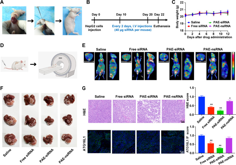
Antitumor effects of PAE-siRNA complex in orthotopic model. A Scheme of the establishment of orthotopic tumor model. HepG2 cells were injected into the lower lobe of liver of Balb/c nude mice to form orthotopic tumors. B Schematic illustration of orthotopic model establishment and treatment. C Changes of mouse body weight during experimental period. Data are shown as means ± SD (n = 6). D Schematic cartoon of PET/CT detection in mice. E Representative images of PET/CT scans from each group. Images of mice were acquired 24 h after intravenous injection of 18F-FDG. Shown from left to right are the axial, coronal, and lateral views. The white circles and red arrows indicated the regions of hepatic tumors. Standardized uptake values (SUV) were examined by 18F-FDG. F Representative images of the HepG2 orthotopic HCC tumors. The white dotted lines indicate tumor regions (n = 6). G Representative images (left) and quantification (right) of liver tumor sections stained with H&E (top) and immunofluorescence (bottom) of ATG16L1. *p < 0.05; **p < 0.01. Scale bars are 50 µm

