Fig. 3.
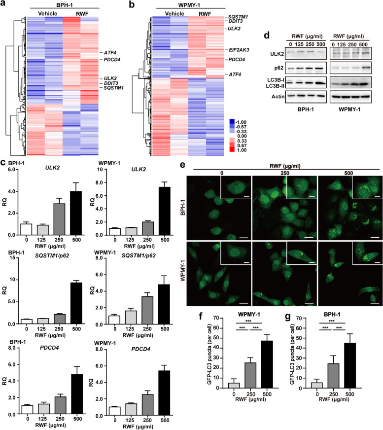
RWF extract regulated autophagy pathway in BPH-1 and WPMY-1 cells. a, b The expression profiling analysis of BPH-1 and WPMY-1 cells exposed to RWF extract (250 μg/ml) or vehicle for 24 h by microarray. The expression levels of autophagy-related genes (ULK2 and SQSTM1/p62) and ER-stress-associated gene (EIF2AK3/PERK, ATF4, and DDIT3/CHOP) were indicated. c, d The mRNA and protein levels of autophagy-associated genes in BPH-1 and WPMY-1 cells treated with RWF extract for 24 h were detected by qRT-PCR (c) and immunoblotting (d) assays, respectively. e BPH-1 and WPMY-1 cells with the stable expression of GFP-LC3 were treated with RWF (250 or 500 μg/ml) or vehicle for 24 h. GFP-LC3 puncta patterns were examined under a LEICA confocal microscope. Scale bars: 25 μm; inset, 10 μm. f, g Quantification of the number of LC3-GFP puncta per cell in BPH-1 (f) and WPMY-1 cells (g). 30 individual cells were counted in each group. ***p < 0.001

