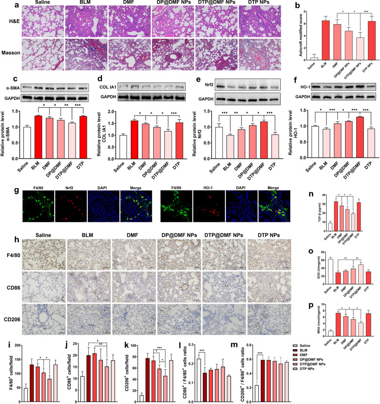
Fig. 7.
DTP@DMF NPs suppress fibrosis and macrophage accumulation in lung tissue via Nrf2 signaling. a Representative H&E and Masson’s trichrome staining and b the severity score of fibrosis after DMF, DP@DMF NPs and DTP@DMF NPs treatment (n = 5). c α-SMA and d collagen Ia1 protein levels in fibrotic tissue (n = 3). e Nrf2 and f HO-1 levels in fibrotic tissue (n = 3). g The Nrf2 (red) and HO-1 (red) expression in F4/80+ macrophages (green) detected by immunofluorescence analysis. h Immunohistochemistry of F4/80+, CD86+, and CD206+ macrophages in pulmonary tissues, and i–m quantification of total macrophages and M1 and M2 phenotypes (n = 5). n TGF-β levels in BALF, o SOD and p MDA levels in tissue (n = 5). Statistical analyses were performed via one-way ANOVA with S–N-K post hoc analysis. * P < 0.05, ** P < 0.01, *** P < 0.001

