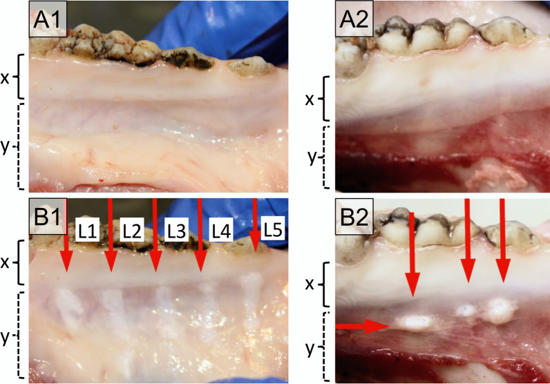
Fig. 6.
Pictures of the oral mucosa of the mandibula of pig cadavers were treated with the Dental water jet (left column, A1–B1) and a powder jet device (right column, A2–B2). Upper panel shows the tissue before (A) and the lower panel after treatment (B) in the region of keratinised gingiva tissue (x) and non-keratinised gingiva (y). The Dental water jet was used with 5 different power settings (L1–L5). Three different areas were tested for the powder jet device. No abrasion is seen in the keratinised mucosa near the teeth (x-range), tissue abrasion and blistering are observed in the non-keratinised mucosa (y-range)

