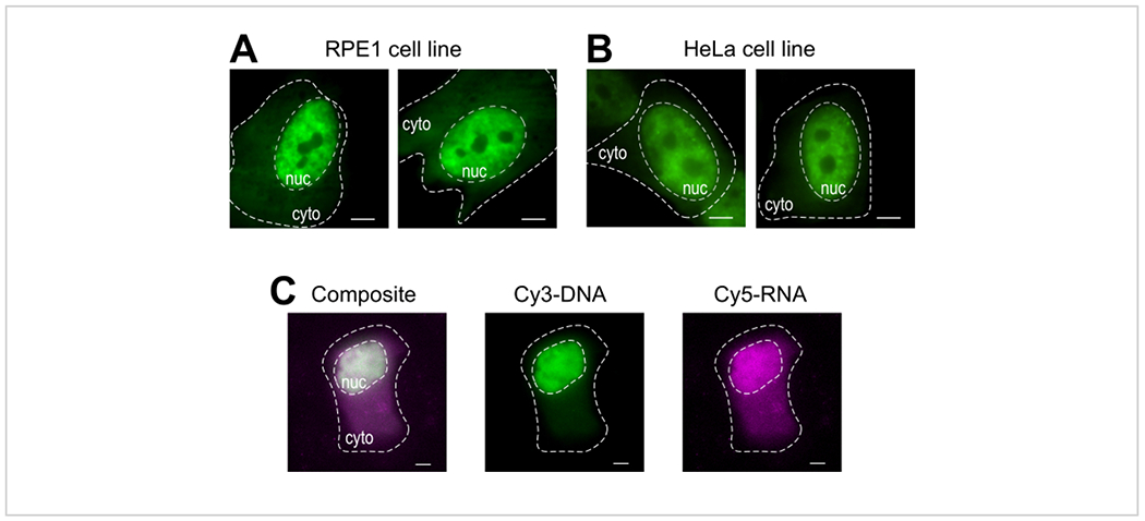
Figure 4: Variations in cell type loading material of the bead loading protocol.

(A-B) RPE1 (A) and HeLa (B) cells were bead-loaded with 1.5 μg of a nuclear Fab protein (anti-RNAPII-Serine 5-phosphorylation) in 4 μL of loading solution. Each cell’s nucleus (nuc) and cytoplasm (cyto) are marked. Cells were imaged 6 h after being bead-loaded. Scale bars = 5 μm. (C) Human U2OS cells were bead-loaded with both Cy5-RNA 9mer (magenta) and Cy3-DNA 28mer (green) oligos, 10 picomoles of each, in 4 μL of bead loading solution. Cells were imaged 4 h after being bead-loaded. All cell nuclei are highlighted by a dashed line. Scale bars = 5 μm. Abbreviations: RNAPII = RNA polymerase II. Please click here to view a larger version of this figure.
