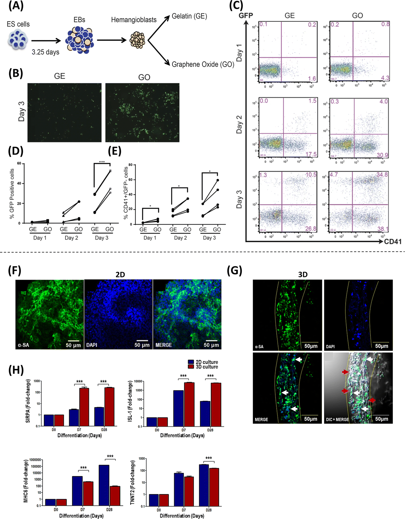
Figure 2. Approaches employing graphene oxide nanosheets and nanofibers to regulate stem cell behavior.

(A) Schematic representation of the experimental design to evaluate the response of hemangioblasts to gelatin (GE) or graphene oxide (GO). Heaemanglioblast cultures on GE/GO showing higher (B and D) GFP signal and (C and E) production of CD41+ / GFP− cells on GO-coated coverslips compared to GE (Adapted from Garcia-Alegria et al., 2016. Ref 14). Expression of sarcomeric alpha-actinin (α-SA), a cardiomyocyte marker, in human iPSCs (hiPSCs) differentiated using gelatin-PCL nanofiber scaffolds in a (F) 2D and (G) 3D culture system. (H) Gene expression of cardiac progenitor and cardiomyocyte markers SIRPA/ISL1, MHC6/TNNT2, respectively during differentiation of hiPSCs into cardiomyocytes in 2D and 3D cultures (Adapted from Sridharan et al., 2021. Ref 27).
