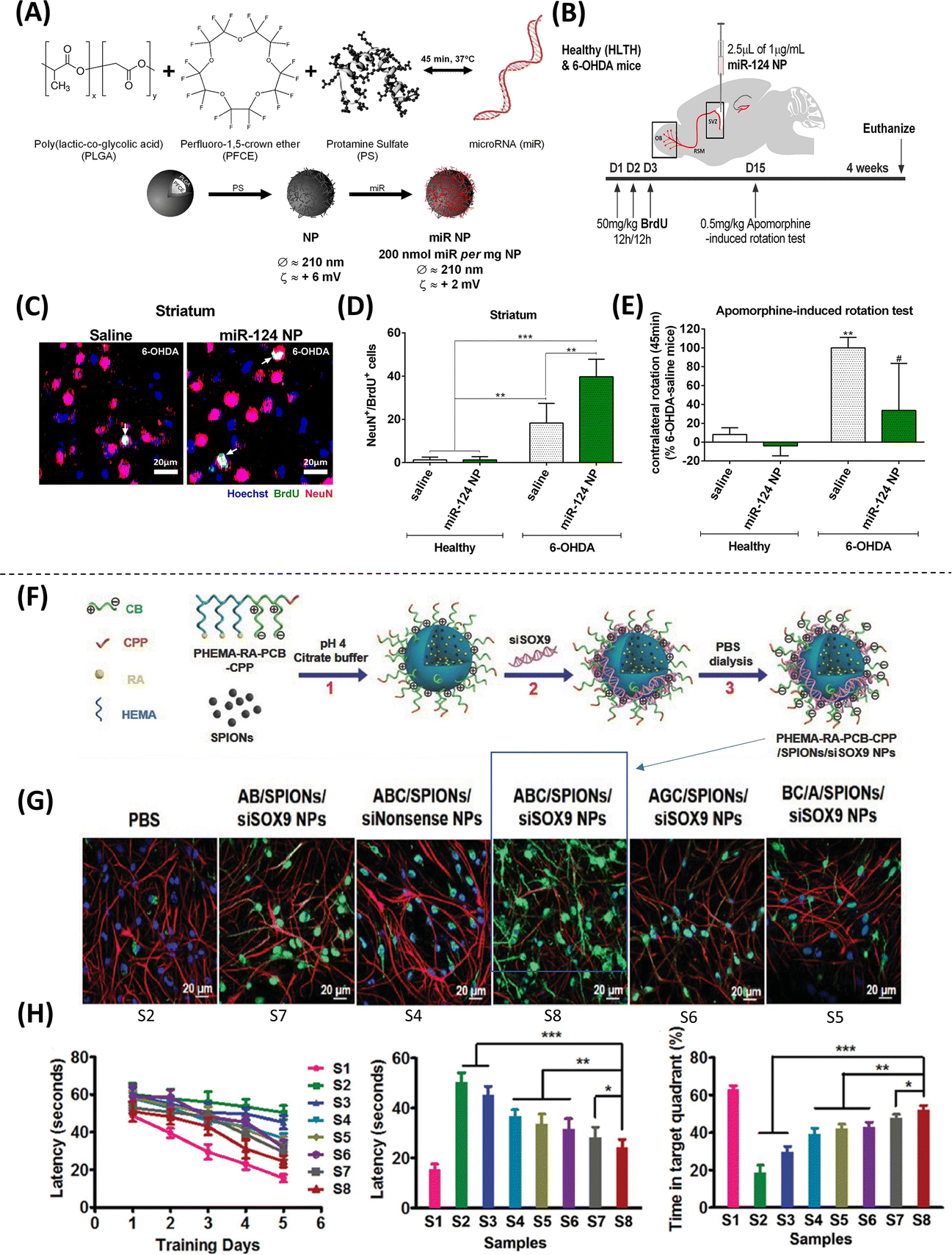
Figure 3. Methodologies using nanoparticles to modulate stem cell differentiation.

(A) Properties of nanoparticles (NPs) loaded with microRNAs. (B) Experimental setup where mice were subjected to two stereotaxic injections, one in the right striatum to deliver 6-Hydroxydopamine (6-OHDA) to induce a PD phenotype, and another in the right lateral ventricle to deliver miR-124 NPs or saline solution. (C-D) Confocal images of BrdU (proliferation marker, green), Hoechst (nuclear marker, blue), and DCX (mature neuronal marker, red) staining showing an increase in the number of mature neurons (NeuN+/BrdU+ cells) observed in the striatum of mice treated with 6-OHDA and miR-124 NPs compared to healthy controls. (E) Apomorphine-rotation test (behavioral analysis) illustrates a decrease in motor deficits (net contralateral rotations) in mice treated with miR-124 NP (Adapted from Saraiva et al., 2016. Ref 37). (F) Composition of the traceable NPs PHEMA-RA-PCB-CPP/SPIONs/siSOX9 (condensed as ABC/SPIONs/siSOX9 NPs: S8). (G) Immunostaining analysis with MAP-2 (neuronal marker, green), GFAP (glial cell marker, red), and DAPI (nuclear marker, blue), showing higher MAP-2 expression (conversion into neurons) when treated with S8 compared to control. (H) Morris water maze experiments were performed to assess the effect of NP treatment on spatial learning and memory improvement, showing that NSCs treated with S8 NPs could potentially improve cognition and memory (Adapted from Zhang et al., 2016. Ref 40).
