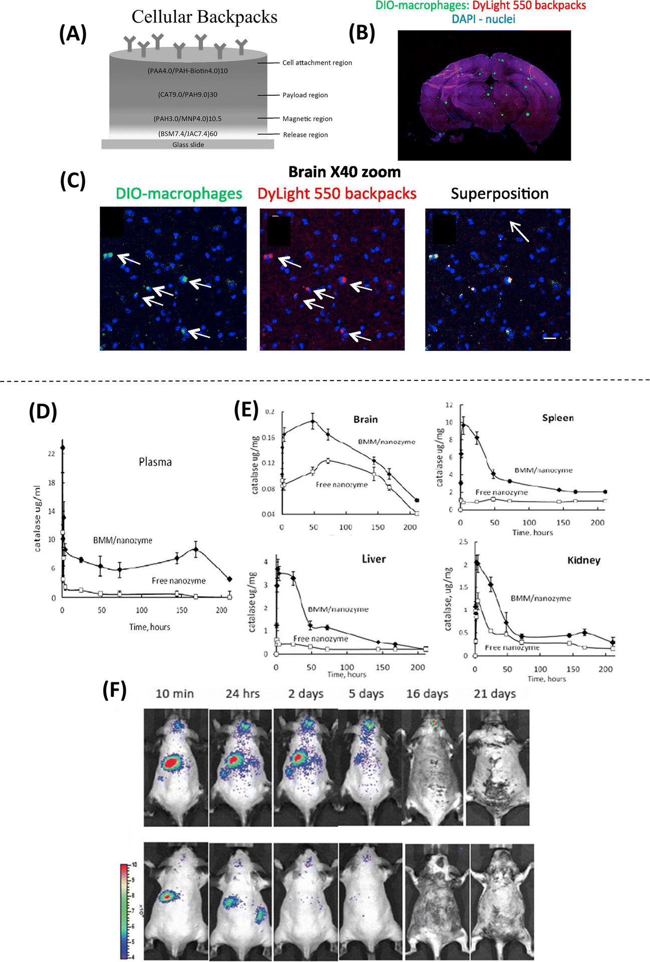
Figure 5. Methodologies employed for cell-mediated drug delivery.

(A) Schematic of the structure of the cellular backpack, loaded with catalase, showing the composition and assembly of different regions from the “release region” to the cell attachment region. (B) Confocal microscopy images of the whole brain after systemic delivery of backpack-carrying macrophages show fluorescently labeled macrophages (green) and backpacks (red). (C) At 40X magnification, co-localization of green and red can be observed, suggesting that the cells facilitated the transport of backpacks to the brain (Adapted from Klyachko et al., 2017. Ref 53). (D) Systemic delivery of bone marrow-derived macrophages (BMM) loaded with a catalase nanozyme in a murine model of brain inflammation shows an increased blood concentration of nanozyme for more than 170 hours after injection. (E) Increased accumulation of catalase is found in all tissues (brain, spleen, liver, and Kidney) when using nanozyme-loaded BMM. Higher accumulation of the nanozyme is found in the spleen and lower accumulation in the brain. (F) Biodistribution of BMM loaded with fluorescently labeled nanozyme, showing targeted drug delivery from peripheral organs to the brain with inflammation for over 16–20 days (top panel) compared to healthy animals (bottom panel) (Adapted from Zhao et al., 2011. Ref 54).
