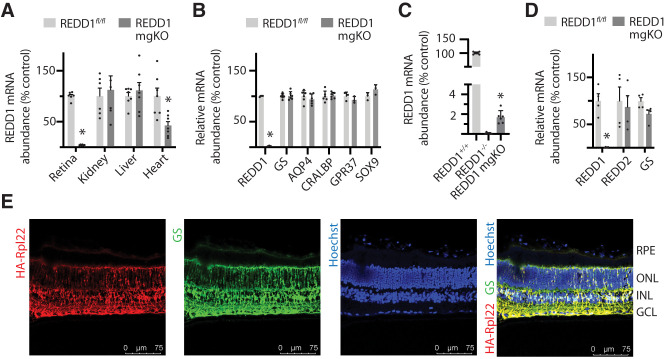
Figure 2.
Conditional deletion of REDD1 nearly eliminated retinal REDD1 expression. A: REDD1 mRNA abundance was evaluated in retina, kidney, liver, and heart of REDD1fl/fl and REDD1-mgKO mice by PCR analysis. B: REDD1, GS, AQP4, CRALBP, GRP37, and SOX9 mRNA abundances were evaluated in retinal lysates. C: REDD1 mRNA abundance was evaluated in the retina of REDD1+/+, REDD1−/−, and REDD1-mgKO mice. D: REDD1, REDD2, and GS expression in primary retinal Müller cells was evaluated. Data points represent biological replicates. Bars are means ± SD (n = 4–7). *P ≤ 0.05 vs. REDD1fl/fl in A, B, and D or REDD1−/− in C. E: Müller cell–specific Cre expression was assessed by crossing Pdgfra-Cre mice with mice homozygous for a mutation targeted to the Rpl22 locus harboring a loxP-flanked wild-type C-terminal exon 4, followed by an identical C-terminal exon 4 that is tagged with three copies of the hemagglutinin (HA) epitope before the stop codon. Whole eyes were isolated and cryo-sectioned into sagittally oriented longitudinal cross-sections. HA-Rpl22 reporter colocalization with the Müller cell–specific marker GS was evaluated in retinal cross-sections by immunofluorescence. Hoechst was used to visualize nuclei. GCL, ganglion cell layer; INL, inner nuclear layer; ONL, outer nuclear layer; RPE, retinal pigment epithelium.

