Figure 3.
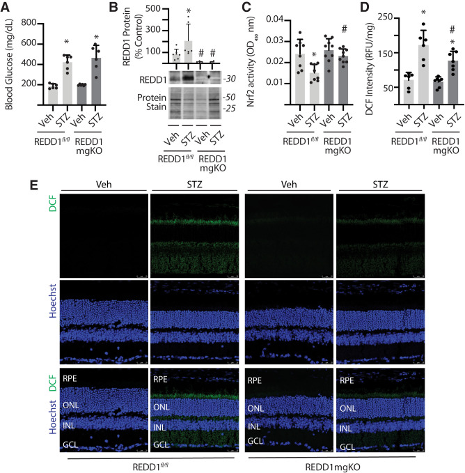
Müller cell–specific REDD1 deletion prevented diabetes-induced Nrf2 suppression and attenuated oxidative stress in the retina. Diabetes was induced in REDD1fl/fl and REDD1-mgKO mice by administration of STZ. Control mice received a vehicle (Veh). All retinal analysis was performed after 6 weeks of diabetes. A: Postprandial blood glucose concentrations were measured. B: REDD1 protein expression was evaluated in retinal lysates by Western blotting. Protein loading was evaluated by reversible protein stain. Blots shown are representative of two independent experiments. Protein molecular mass (kDa) is indicated at right of blots. C: Nrf2 activity was evaluated in the nuclear fraction obtained from retinal lysates of REDD1fl/fl and REDD1-mgKO mice by DNA-binding ELISA. OD, optical density. D: ROS were quantified in the supernatants from whole retinal lysates using DCF. Values are means ± SD (n = 5–9). RFU, relative fluorescent units. Significance was defined as P < 0.05 for all analyses. *P ≤ 0.05 vs. Veh; #P ≤ 0.05 vs. REDD1fl/fl. E: ROS were visualized in whole-eye sagittal cross-sections using DCF. Hoechst was used to visualize nuclei. GCL, ganglion cell layer; INL, inner nuclear layer; ONL, outer nuclear layer; RPE, retinal pigment epithelium.

