Figure 5.
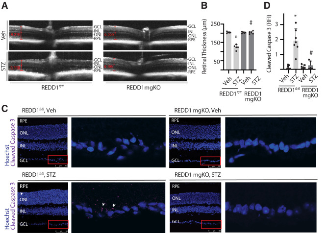
Retinal thinning was not observed in diabetic mice with Müller cell–specific REDD1 deletion. Diabetes was induced in REDD1fl/fl and REDD1-mgKO mice by administration of STZ. Control mice received a vehicle (Veh). All retinal analysis was performed after 6 weeks of diabetes. A: Retinal thickness was evaluated by SD-OCT. Representative images are shown. B: Retinal thickness from the RNFL to the photoreceptor OS was manually measured as indicated by red calipers. C: Whole eyes were isolated and cryosectioned into sagittally oriented longitudinal cross-sections, and cleaved caspase-3 was evaluated by immunohistochemistry. The red box indicates the area presented at higher magnification. White arrowheads indicate cleaved caspase-3–positive immunoreactivity. D: Cleaved caspase-3 intensity was quantified in retinal cross-sections. Bars are means ± SD (n = 5–7). *P ≤ 0.05 vs. Veh; #P ≤ 0.05 vs REDD1fl/fl. INL, inner nuclear layer; ONL, outer nuclear layer; RPE, retinal pigment epithelium; RFI, relative fluorescent intensity.

