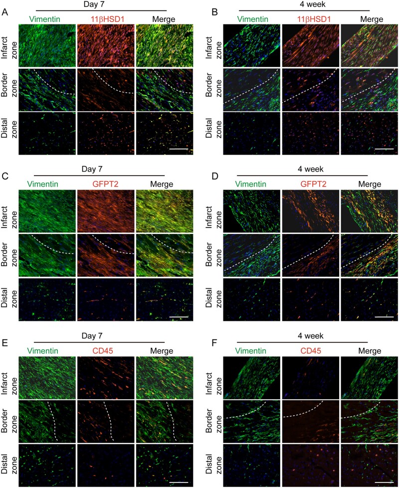
Figure 7.
Expression of Hsd11b1 and Gfpt2 in cardiac fibroblast upon myocardial injury. (A,B) Representative IHC images showing the expression of Hsd11b1 and its colocalization with Vimentin in infarcted region, border zone and distal zone of heart 7 days (A) and 4 weeks (B) post-MI. (C,D) Representative IHC images showing the expression of Gfpt2 and its colocalization with Vimentin in infarcted region, border zone and distal zone of heart 7 days (C) and 4 weeks (D) post-MI. (E,F) Representative IHC images showing the expression of Gfpt2 expression and its colocalization with Vimentin in infarcted region, border zone and distal zone of heart 7 days (E) and 4 weeks (F) post-MI. Dashed lines indicate the borderline between injured and uninjured or less injured area. The experiment was performed with n = 5 mice per each group. In each heart, n = 4 sections at different layers of heart were used.

