Figure 6.

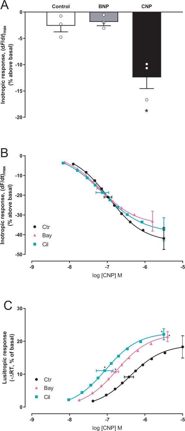
Inhibiting PDE2 or PDE3 sensitizes the CNP-induced lusitropic response. (A) Left ventricular muscle strips were incubated with vehicle (control) or the indicated ligand for 20–25 minutes and maximal inotropic response recorded and shown as per cent relative to that prior to stimulation (basal) (n = 3 strips from three animals, CNP; 100 nM, BNP; 100 nM). (B, C) Concentration–response curves of inotropic response (B) and lusitropic response (C) to CNP in the absence or presence of Bay (Bay60-7550, 50 nM) or Cil (cilostamide, 1 µM). Data are relative to basal (prior to CNP stimulation; n = 6–9). Left ventricular strips were pre-incubated with the indicated PDE inhibitor for 45 min before the first addition of CNP. Horizontal error bars depict SEM of the EC50, and vertical error bars are SEM of the maximal inotropic (B) or lusitropic (C) responses. *P < 0.05 vs. CNP alone (one-way ANOVA with Dunnett’s multiple comparisons test).
