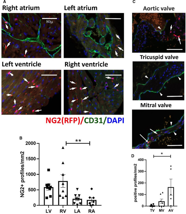
Figure 1. The neuron‐glial antigen 2 (NG2)DsRed reporter model identifies a large population of pericytes in the mouse myocardium.

A, Dual immunofluorescence for red fluorescent protein (RFP) and CD31 identifies NG2+ pericytes in atria and ventricles (arrows), associated with CD31+ endothelial cells (A). B, Quantitative analysis shows that the density of pericytes is higher in the right ventricle (RV) than in the right atrium (RA). Moreover, there is a trend (P=0.08) toward a higher pericyte density in the left ventricle (LV), in comparison to the left atrium (LA). C, Dual immunofluorescence identifies NG2+ cells (arrows) in the valves. The arrowheads indicate the CD31+ valvular endothelial cells. D, The aortic valve (AV) has a higher density of pericytes than the mitral valve (MV) and tricuspid valve (TV) (*P<0.05, **P<0.01; n=5–8 per group). Scalebar=80 μm. Data are expressed as mean±SE. Statistical comparison was performed by ANOVA, followed by the Sidak post hoc test (B), or the nonparametric Kruskal‐Wallis test (D). DAPI indicates 4’,6’‐diamidino‐2‐phenylindole; and Dsred, Discosoma species red.
