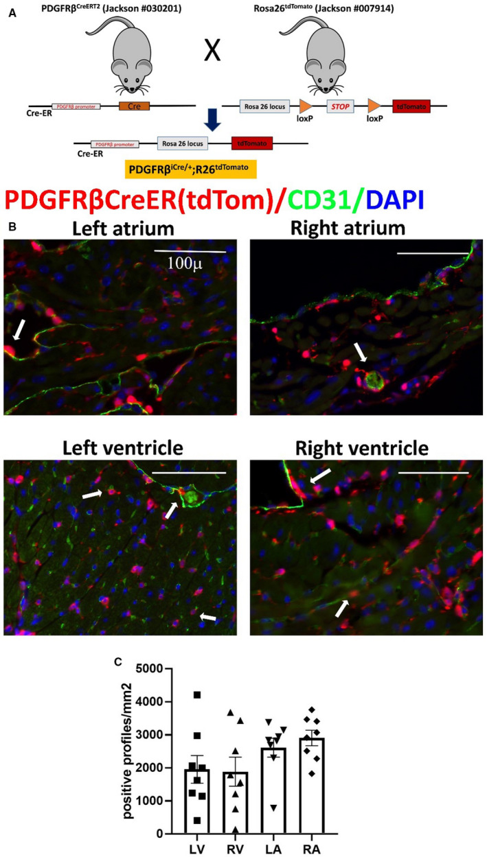
Figure 6. The inducible platelet‐derived growth factor receptor (PDGFR)βCreERT2 driver labels not only periendothelial cells but also a large population of myocardial interstitial cells, which are not associated with vessels.

A, Breeding strategy shows the development of PDGFRβiCre/+;R26tdTomato model. B, Abundant PDGFRβ+ cells are identified in all cardiac chambers using the lineage tracing approach. Please note that the density of the PDGFRβ+ cells is much higher than the numbers of neuron‐glial antigen 2 (NG2)+ cells (shown in Figure 5). Dual fluorescence with CD31 shows that a significant proportion of these cells is not associated with endothelial cells. C, Quantitative analysis (n=8) of the density of PDGFRβ+ cells in atria and ventricles. Scalebar=100 µm. Data are expressed as mean±SE. Statistical comparison was performed using nonparametric ANOVA (Kruskal‐Wallis, P=not significant). LA indicates left atrium; LV, left ventricle; RA, right atrium; RV, right ventricle; and tdTom, tandem dimer Tomato.
