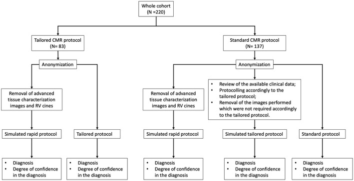
Figure 2. Assessment of advanced tissue characterization value.

Scans in the tailored CMR group had been anonymized and reported twice (ie, with and without advanced tissue characterization). Scans in the control group (standard CMR protocol) had been anonymized and reported 3 times: (1) without any advanced tissue characterization image or RV cine images, simulating the rapid protocol of the INCA Peru Study 9 ; (2) with advanced tissue characterization and cine images accordingly to the tailored CMR approach; and (3) with all the original images. Each time, the reporting physician expressed a diagnosis and a degree of confidence in that diagnosis (poor, moderate, strong). CMR indicates cardiac magnetic resonance; and RV, right ventricle.
