
Figure 1. Echocardiographic visualization of mitral valve prolapse (MVP) and associated left ventricular (LV) fibrosis.
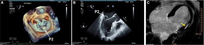
A and B, Three‐dimensional and 2‐dimensional transesophageal echocardiography MVP showing prolapse of the P2 segment (white arrows). C, Cardiac magnetic resonance with late gadolinium enhancement showing evidence of inferobasal LV fibrosis underlying the papillary muscles (yellow arrows).