
Figure 2. Regional left ventricular (LV) fibrosis in human patients with mitral valve prolapse (MVP).
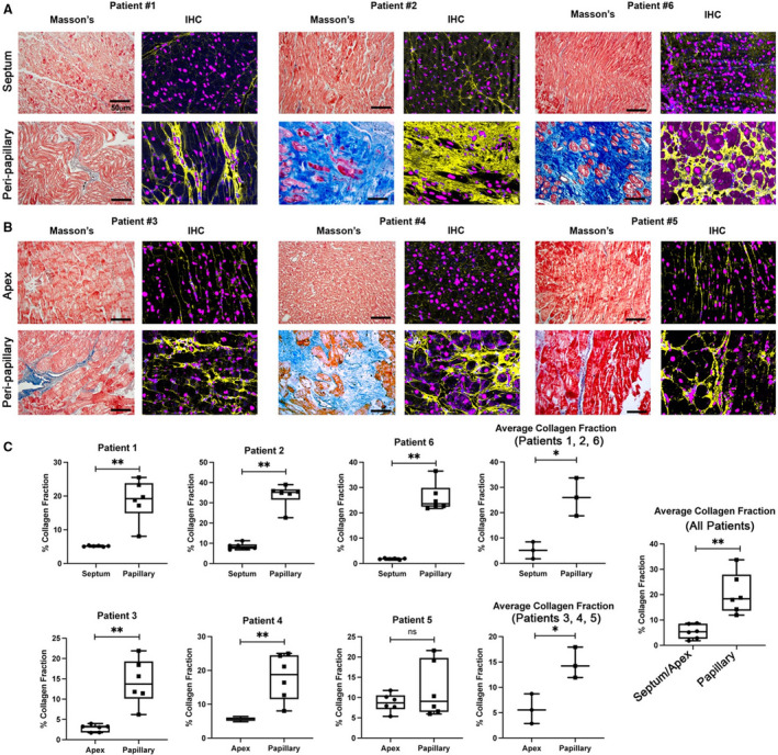
Masson’s trichrome and immunohistochemistry (IHC) for collagen (yellow) shows prominent LV fibrosis in the peripapillary region of surgical mitral valve repair patients compared with either septal (A) or apex (B) biopsies. C, Quantification of fibrosis shows significant elevation of collagen I protein in peripapillary regions compared with either septal or apex in‐person control tissue. Amounts are shown as percent collagen fraction (positive pixel staining/total pixels). Zones of myocyte loss are evident, indicating replacement fibrosis. Scale bars=50 µm. Blue=collagen histological (Masson) stain, red=myocytes histological (Masson) stain, purple=nuclei (Hoechst). *P<0.05, **P<0.01.