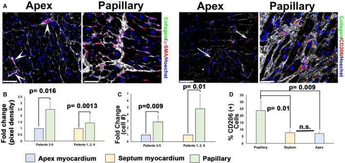
Figure 3. Fibrosis in patients with mitral valve repair correlates with activated cell types specific to peripapillary regions.

A, Immunohistochemistry (IHC) (white arrows, purple staining) shows increased α‐smooth muscle actin (α‐SMA) and cluster of differentiation 206 positive (CD206+) macrophages within fibrotic areas (white, collagen) localized to peripapillary regions (arrows) of the left ventricle compared with in‐person apex tissue. Scant α‐SMA+ cells within capillaries are observed in apex tissue (arrowheads in A), and few macrophages are present within the apex (arrows). B through D, Quantification of IHC data shows 2‐ to 3‐fold increase in α‐SMA expression, a 3‐ to 5‐fold increase in cluster of differentiation 206 (CD206) macrophages, and ≈25% total CD206+ cells within the fibrotic peripapillary region compared with apex or septal in‐person control tissues. Scale bars=100 µm.
