
Figure 5. Regionalized fibrosis progression is observed in Dzip1S14R/+ mice.
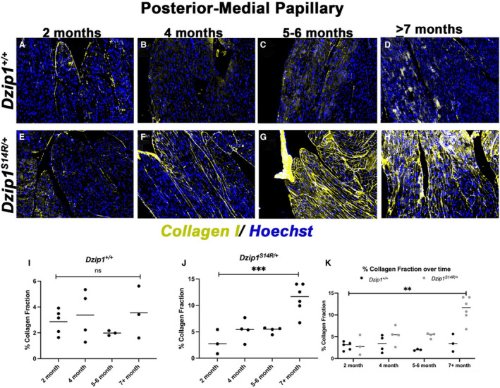
A through D, Immunohistochemistry for collagen I (yellow) over time shows no discernible increase of collagen production within the posterior–medial papillary muscle of control mice (Dzip1+/+ ). E through H, Dzip1S14R/+ mitral valve prolapse mice show increased collagen I staining (yellow) over time within the papillary muscle. I, Quantification of percent collagen fraction of tissue within the control papillary muscle shows no significant change in expression over time. One‐way ANOVA resulted in a P=0.55. J, Compared with 2 months of age, the Dzip1S14R/+ posterior–medial papillary muscle shows a trend toward increased fibrosis by 5 to 6 months of age and ≈5‐fold increase by ≥7 months. ***One‐way ANOVA resulted in a P=0.0004. K, Compared with control animals, significant differences in collagen within the papillary muscle is observed in Dzip1S14R/+ mice (2‐way ANOVA P for genotype=0.0005, P for time point<0.0001, P for interaction=0.004). Post hoc comparison of individual time points using Bonferroni multiple comparisons test found that significant differences in collagen are present by ≥7 months of age (P<0.0001), whereas a trend for increased collagen is observed by 6 months of age (P=0.12). ns, not significant; **P<0.01; ***P<0.001.