
Figure 4. Left ventricular longitudinal hemodynamic force (basal‐apical): diastolic phase.
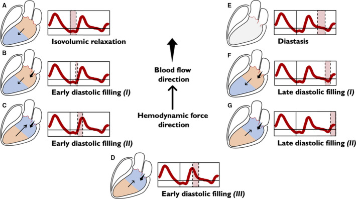
The intraventricular pressure gradients are illustrated for simplicity through only 2 areas: the area at a higher pressure is shown in orange and the area at a lower pressure is shown in blue. The absence of intraventricular gradient is colored in gray. The direction of the hemodynamic force (thin arrow) always goes from the higher toward the lower pressure area. The direction of flow (wide arrow) goes from the higher to the lower pressure chamber. Diastole begins with the isovolumic relaxation (A), in which both the aortic and the mitral valves are closed. During this period, there is no flow between the cardiac chambers, but because of active myocardial relaxation and recoil of elastic forces generated during the previous systole, the pressure gradient directed toward the ventricular apex increases, thus generating a diastolic suction before the opening of the mitral valve. This phase persists until the LV pressure drops below the left atrial pressure, the mitral valve opens, and the early diastolic filling begins; ventricular filling at the beginning is passive and the HDF vector continues to be directed toward the LV apex, but the pooling of blood within the LV (toward the apex) rapidly reduces the HDF toward zero (B). After this stage, LV filling continues supported by the upward movement of the mitral plane that displaces the blood contained into the atrium inside the LV. In this phase, gradually, the pressure in the LV increases until it exceeds the atrial pressure, thus inverting the A‐V pressure gradient, decelerating the LV filling and making HDF to grow in the positive ascending phase (C). The reduced passage of blood from the atrium to the LV progressively equilibrates the pressures in both chambers, eventually reducing the gradient to zero, causing a positive descending phase on the HDF curve (D). In the next phase (diastasis), a pressure equilibrium is established between the base and apex (and between the ventricle and left atrium) (E). The occurrence of atrial contraction, causes a relative gradient from apex to base, resulting in HDF negative vectors (F) and producing the late diastolic filling. Once again, as blood accumulates in LV, the ventricular gradient is reversed and HDF vector become positive (G), decelerating the diastolic filling flow and preparing LV for the systolic ejection phase. A‐V indicates atrio‐ventricular; HDF, hemodynamic force; and LV, left ventricle.