
Figure 5. Relationship between heartbeat events and longitudinal hemodynamic force.
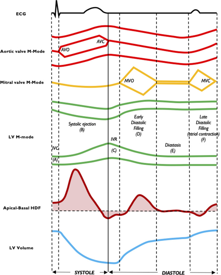
Time coupling among apical‐basal HDF, LV volume, mitral and aortic valve M‐mode, and LV M‐mode is shown. Isovolumic contraction (A), systolic ejection (B), isovolumic relaxation (C), early diastolic filling (D), diastasis (E) and late diastolic filling (F). AVC indicates aortic valve closure; AVO, aortic valve opening; ECG, electrocardiography; HDF, hemodynamic force; IVC, isovolumic contraction; IVR, isovolumic relaxation; LV, left ventricular; MVC, mitral valve closure; and MVO, mitral valve opening.