
Figure 8. Left ventricular longitudinal hemodynamic force (apical‐basal) in normal and pathologic hearts.
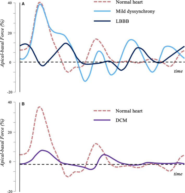
A, Longitudinal HDF in normal heart (red), in mild dyssynchrony with preserved EF (light blue) and in LBBB with reduced EF (dark blue) are shown. Mild dyssynchrony is associated with a modulation of the systolic wave, due to the asynchronous contraction of different regions, and a rebound effect during the diastolic relaxation phase. In presence of LBBB, the asynchrony is even more evident, especially in systole. B, Longitudinal HDF in normal heart (red) and in DCM with reduced EF (purple) are shown. In absence of asynchrony, DCM presents a generalized reduction in HDF amplitude. DCM indicates dilated cardiomyopathy; EF, ejection fraction; HDF, hemodynamic force; and LBBB, left bundle branch block.