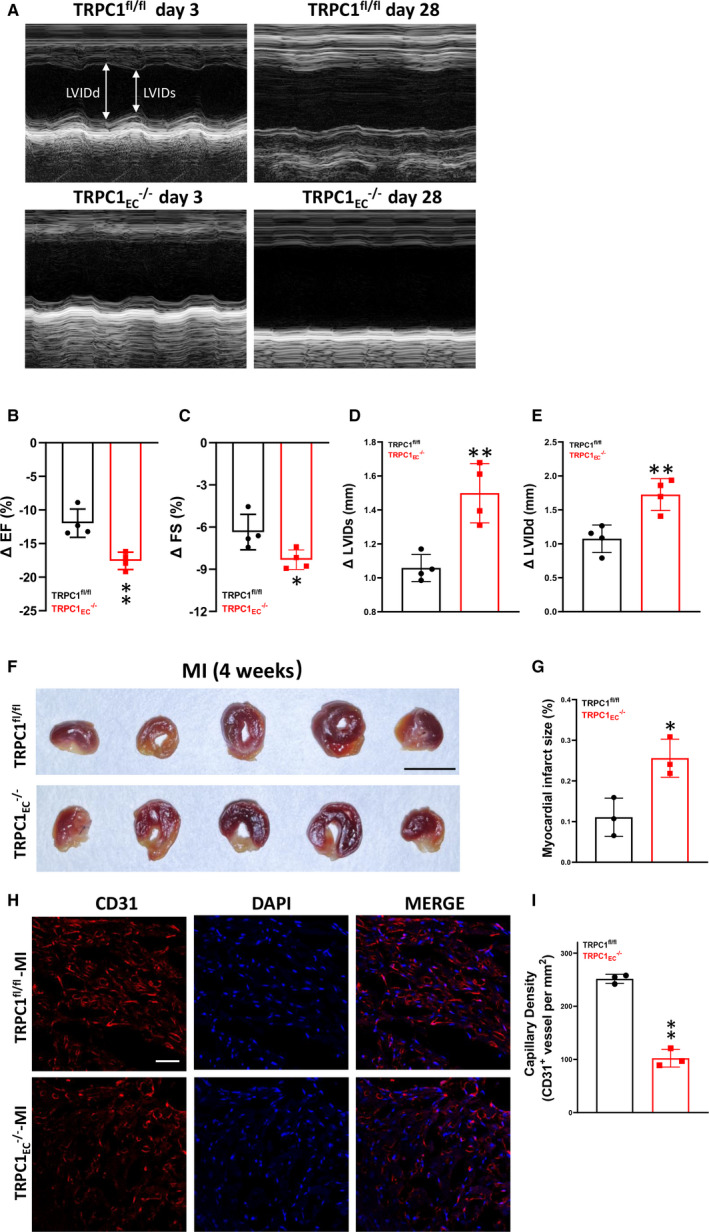
Figure 2. Effects of transient receptor potential canonical 1 (TRPC1) on angiogenesis in myocardial infarction‐model mice.

A, Representative echocardiogram of coronary artery endothelial cells from floxed TRPC1 (TRPC1fl/fl) and TRPC1EC ‐/‐ mice at 3 days and 28 days post‐myocardial infarction. B, Change in ejection fraction from 3 days to 28 days after left anterior descending artery ligation in TRPC1fl/fl and TRPC1EC ‐/‐ mice. C, Quantitative analysis of fractional shortening. D, Quantitative analysis of systolic left ventricular internal dimensions. E, Quantitative analysis of diastolic left ventricular internal dimensions. (n=4; ** P<0.01 vs TRPC1fl/fl, unpaired t test). F, Infarct size in TRPC1fl/fl and TRPC1EC ‐/‐ groups measured by triphenyltetrazolium chloride staining (scale bar, 0.5 cm). G, Statistics of the relative infarct size (n=3; * P<0.05 vs TRPC1fl/fl, unpaired t test). H, Representative confocal laser scanning microscope images showing capillary density in hearts from TRPC1fl/fl and TRPC1EC ‐/‐ myocardial infarction‐model mice (red, cardiac CD31; blue, DAPI; scale bar, 20 μm). I, Quantitation of capillary density in remote zone of left ventricle from TRPC1fl/fl and TRPC1EC ‐/‐ myocardial infarction‐model mice (n=3; * P<0.05 vs TRPC1fl/fl, unpaired t test). The data are presented as mean ± SD. DAPI indicates 4’,6‐diamidino‐2‐phenylindole; EF, ejection fraction; FS, fractional shortening; LVIDd, diastolic left ventricular internal dimensions; LVIDs, systolic left ventricular internal dimensions; MI, myocardial infarction; TRPC1EC ‐/‐, endothelial cell‐specific TRPC1 channel knockout; TRPC1fl/fl, coronary artery endothelial cells from floxed TRPC1; and siTRPC, TRPC siRNA.
