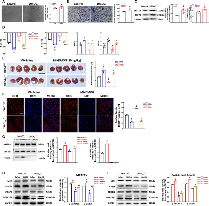
Figure 4. Upregulation of transient receptor potential canonical 1 (TRPC1) by dimethyloxalylglycine (an HIF‐1α [hypoxic inducible factor‐1α] activator) promotes recovery from ischemia.

A, Representative images and quantification of tube formation in primary wild‐type mouse coronary artery endothelial cells (MCAECs) treated with saline or dimethyloxaloylglycine (200 μmol/L, 24 hours) (n=5; * P<0.05 vs control, unpaired t test; scale bar, 50 μm). B, Representative images and quantification of migrated wild‐type MCAECs treated with saline or dimethyloxaloylglycine (n=3; *P<0.01 vs control, unpaired t test; scale bar, 50 μm). C, Protein expression levels of HIF‐1α and TRPC1 in wild‐type MCAECs treated with saline or dimethyloxaloylglycine (n=4; ** P<0.01 vs control, unpaired t test). D, Change in ejection fraction, fractional shortening, systolic LV internal dimensions, and diastolic left ventricular internal dimensions from baseline to 28 days after left anterior descending artery ligation in coronary artery endothelial cells from floxed TRPC1 (TRPC1fl/fl) and TRPC1EC ‐/‐ mice treated with saline or dimethyloxaloylglycine (30 mg/kg per day intraperitoneally) (n=3–5; ** P<0.01 vs TRPC1fl/fl + saline, ## P<0.01 vs TRPC1EC ‐/‐ + saline, ++ P<0.01 vs TRPC1fl/fl + dimethyloxaloylglycine, 1‐way ANOVA and Tukey multiple comparisons test). E, Infarct size in TRPC1fl/fl and TRPC1EC ‐/‐ myocardial infarction‐model mice treated with saline or dimethyloxaloylglycine measured by triphenyltetrazolium chloride staining (n=3; * P<0.05 and ** P<0.01 vs TRPC1fl/fl + saline, ## P<0.01 vs TRPC1fl/fl + dimethyloxaloylglycine, 1‐way ANOVA and Tukey multiple comparisons test; scale bar, 0.5 cm). F, Representative confocal laser scanning microscope images and statistics for capillary density in TRPC1fl/fl and TRPC1EC ‐/‐ myocardial infarction‐model mice treated with saline or dimethyloxaloylglycine (red, cardiac CD31; blue, DAPI, scale bar, 20 μm; n=3; ** P<0.01 vs TRPC1fl/fl + saline, ## P<0.01 vs TRPC1fl/fl + dimethyloxaloylglycine, one‐way ANOVA and Tukey multiple comparisons test). G, Protein expression levels of HIF‐1α and TRPC1 in MCAECs of post‐infarct hearts from TRPC1fl/fl and TRPC1EC ‐/‐ myocardial infarction‐model mice treated with saline or dimethyloxaloylglycine (n=3; ** P<0.01 vs TRPC1fl/fl + saline, ## P<0.01 vs TRPC1EC ‐/‐ + saline, ++ P<0.01 vs TRPC1fl/fl + dimethyloxaloylglycine, 1‐way ANOVA and Tukey multiple comparisons test). H, Representative blots and quantitative analysis of Western blot of P‐MEK and P‐ERK in primary TRPC1fl/fl and TRPC1EC ‐/‐ MCAECs treated with saline or dimethyloxaloylglycine (n=3; * P<0.05 and ** P<0.01 vs TRPC1fl/fl + saline, ## P<0.01 vs TRPC1EC ‐/‐ + saline, 1‐way ANOVA and Tukey multiple comparisons test). I, The protein levels of P‐MEK and P‐ERK in post‐infarct hearts from TRPC1fl/fl and TRPC1EC ‐/‐ myocardial infarction‐model mice treated with saline or dimethyloxaloylglycine (n=3; * P<0.05 and ** P<0.01 vs TRPC1fl/fl + saline, ## P<0.01 vs TRPC1EC ‐/‐ + saline, 1‐way ANOVA and Tukey multiple comparisons test). The data are presented as mean ± SD. DAPI, 4',6‐diamidino‐2‐phenylindole; EF, ejection fraction; FS, fractional shortening; HIF‐1α, hypoxic inducible factor‐1α; LVIDd, diastolic left ventricular internal dimensions; LVIDs, systolic left ventricular internal dimensions; MI, myocardial infarction; TRPC1EC ‐/‐, endothelial cell‐specific TRPC1 channel knockout; and TRPC1fl/fl, coronary artery endothelial cells from floxed TRPC1, P‐MEK, phosphorylated mitogen‐activated protein kinase; and P‐ERK, phosphorylatedextracellular signal‐regulated kinase.
