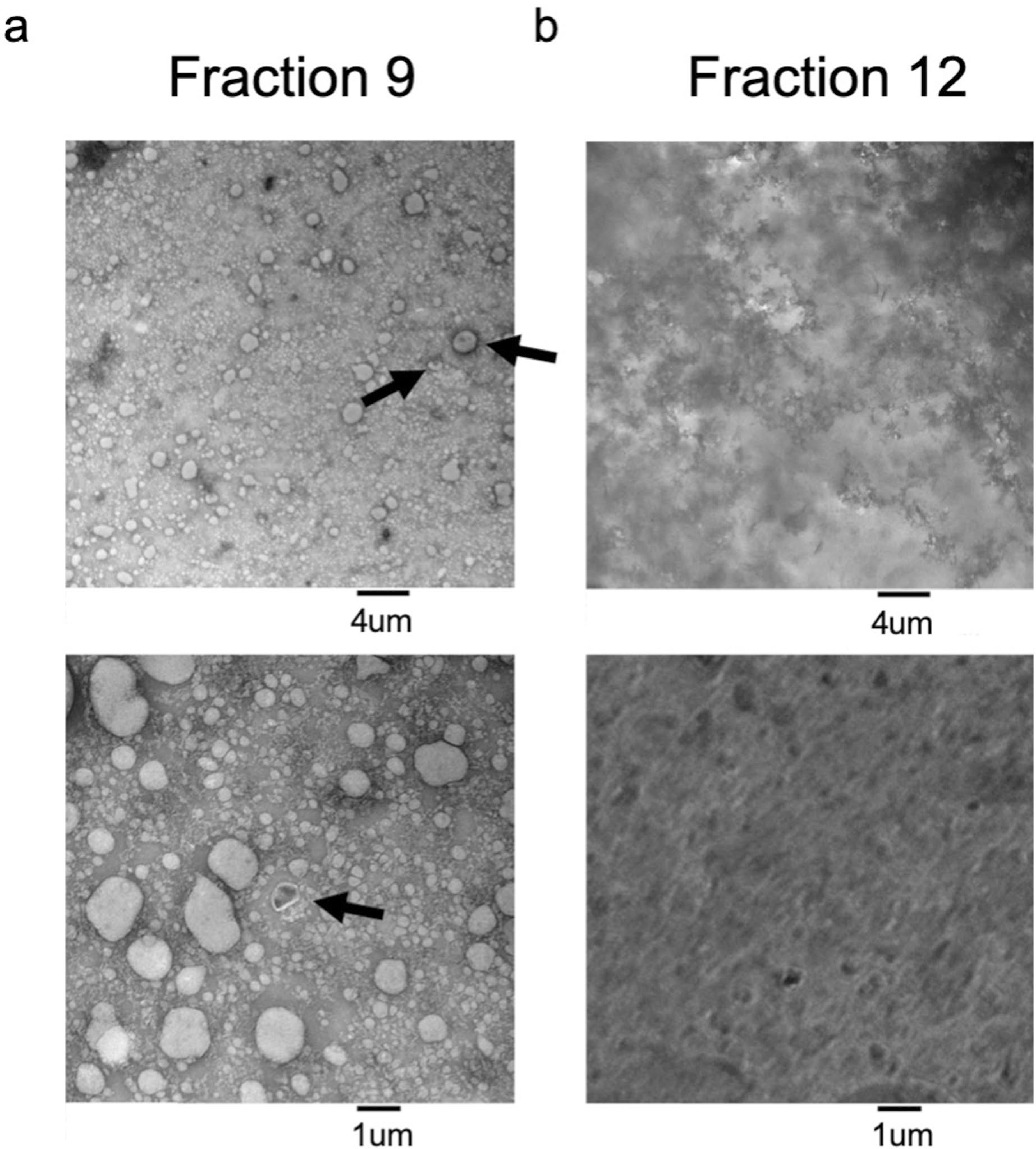
Extended Data Fig. 2 |. Electron Microscopy of SEC fractions.

Transmission Electron Microscopy of a. Fraction 9 and b. Fraction 12 from plasma fractionated using SEC and negatively stained with uranyl formate. Representative images are shown at 6000× magnification (top) and 20,000× magnification (bottom). Arrows indicate ‘cup-shaped’ EVs. This experiment was conducted once.
