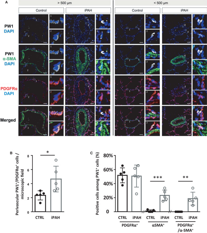
Figure 1. Perivascular PW1+/PDGFRα+ cells are increased in the perivascular zone and within the vascular wall of remodeled arteries in patients with iPAH.

Lung sections from control (CTRL) or patients with idiopathic pulmonary arterial hypertension (iPAH) were labeled for PW1 (white), PDGFRα (red), and α‐SMA (green). A, Representative confocal images of pulmonary vessels (>500 µm diameter, left panels; <500 µm diameter, right panels) from control patients (n=5) and patients with iPAH (n=5). Panels 1, 2, and 3 show details of perivascular PW1+/PDFGFRα+ cells. Panel 4 shows details of PW1+/PDFGFRα+/α‐SMA+ cells found within arteries in patients with iPAH. B, quantification of perivascular PW1+/PDGFRα+ cells. C, quantification of PDGFRα+ or α‐SMA+ or PDGFRα+/α‐SMA+ cells among perivascular and vascular PW1+. Scale bar, 50 µm. Bars represent means and whiskers represent SD. *P<0.05, **P<0.01, ***P<0.001; ns indicates not significant (2‐tailed Mann‐Whitney); PDGFRα indicates platelet‐derived growth factor receptor type α; PW1, protein widely 1; and SMA, smooth muscle actin.
