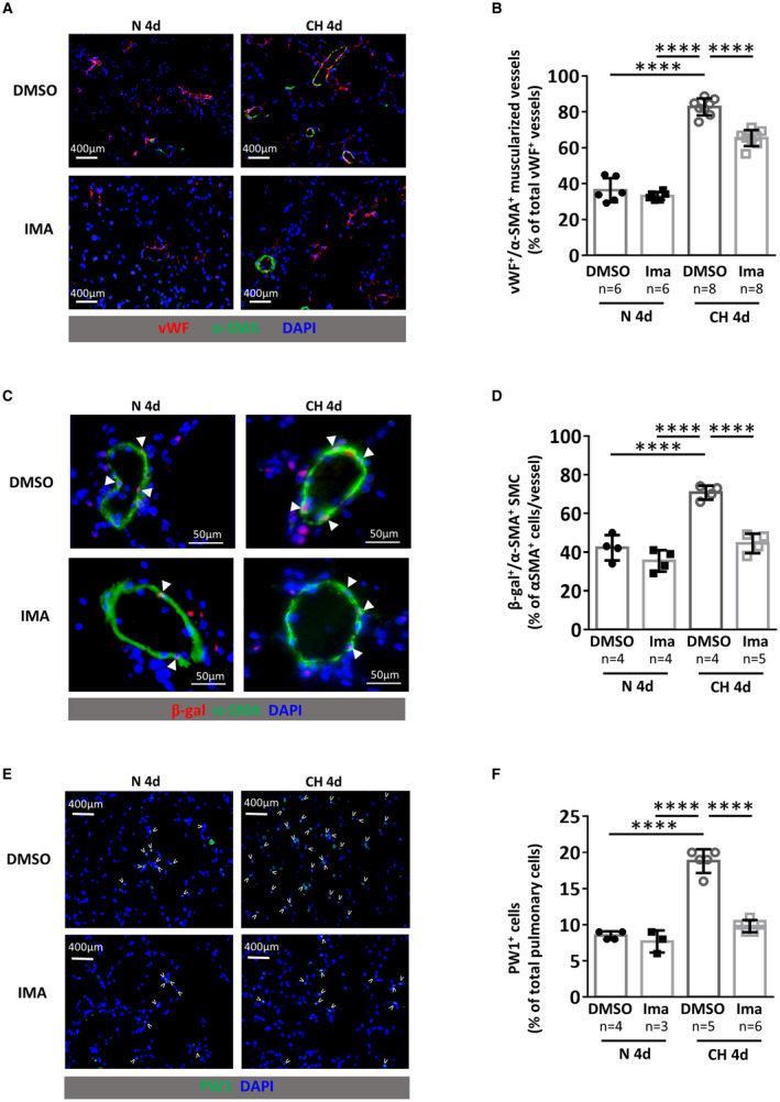
Figure 2. Imatinib treatment prevents early chronic hypoxia (CH)‐induced neomuscularization and PW1+ progenitor cell recruitment and differentiation in SMCs.

PW1nLacz mice were maintained under normoxia (N 4d) or chronic hypoxia (CH 4d) for 4 days and treated daily with DMSO or imatinib (IMA). A, Representative images of von Willebrand factor (vWF, red) and α‐smooth muscle actin (α‐SMA, green) staining in lungs. B, Quantification of muscularized vessels (fully+partially) (n=6–8 mice/group). C Representative images of pulmonary muscularized vessel stained with α‐SMA (green) and β‐galactosidase (red) in lungs (double positive are marked by arrowheads). D, Quantification of lung β‐galactosidase (β‐gal)‐expressing SMC in fully‐muscularized vessels (n=4–5 mice/group). E, Representative images of lung parenchyma stained for PW1 (PW1 in green, positive cells are marked by arrowheads). F, Quantification of PW1+ cells in lung parenchyma (n=3–6 mice/group). Bars represent means and whiskers represent SD. ****P<0.0001, 2‐way ANOVA and Tukey. PW1 indicates protein widely 1; and SMCs, smooth muscle cells.
