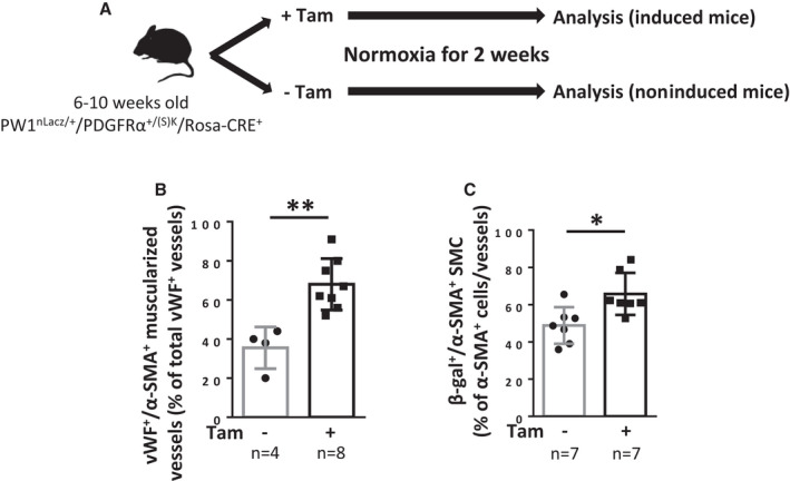
Figure 4. Constitutive PDGFRα activation leads to pulmonary vessel neomuscularization because of increased numbers of PW1+ progenitor‐derived SMCs.

A, Timeline of tamoxifen (Tam) induction and analysis of PW1nLacz/+/PDGFRα+/(S)K/Rosa‐CRE+ mice (males + females). B and C, Analysis of mice lungs 2 weeks after tamoxifen induction. B, Quantification of muscularized vessels in lungs from control or tamoxifen‐treated mice. Pulmonary vessel muscularization was determined by immunofluorescence using anti–α‐smooth muscle actin (α‐SMA) and anti‐von Willebrand factor (vWF) antibodies (n=4–8). For each animal, ≈100 vWF+ vessels (<100 µm) were analyzed for muscularization (α‐SMA+). C, Quantification of lung β‐galactosidase (β‐gal)‐expressing SMC in fully‐muscularized pulmonary vessels. The percentage of lung β‐gal+/α‐SMA+ cells among α‐SMA+ cells in fully‐muscularized vessels (<100 µm) was determined by immunofluorescence (n=7 for each condition). Bars represent means and whiskers represent SD. *P<0.05, **P<0.01 (2‐tailed Mann‐Whitney). PDGFRα indicates platelet‐derived growth factor receptor type α; PW1, protein widely 1; SMA, smooth muscle actin; and SMC, smooth muscle cell.
